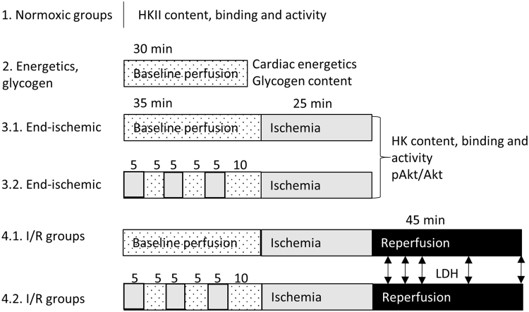
Figure 1.
Schematic overview of the different protocols. All hearts were exposed to 20 min perfusion with either glucose only, or multi-substrate perfusate to stabilize the heart. Hearts of group 1 were then homogenized and mitochondria were isolated for determination of mitochondrial hexokinase amount and activity. Hearts of group 2 were exposed to 30 min baseline perfusion after which they were frozen and used for cardiac energetics and glycogen determination. Hearts of group 3 were exposed to 35 min baseline perfusion with or without IPC, followed by 25 min no-flow ischemia. Then hearts were homogenized and mitochondria isolated for determination of HKII amount and activity and Akt phosphorylation. Hearts of group 4 were exposed to 35 min baseline perfusion with or without IPC, 25 min ischemia and 45 min reperfusion. LDH was sampled at 5, 10, 15, 30 and 45 min reperfusion to determine cardiac damage. Hearts of group 5 were exposed to 20 min baseline perfusion with multi-substrate buffer, followed by 25 min ischemia and 10 min reperfusion. White: baseline perfusion with multi-substrate perfusate, white dotted: baseline perfusion with both glucose-only and multi-substrate perfusate, grey: no-flow ischemia, black: reperfusion.

