Figure 7.
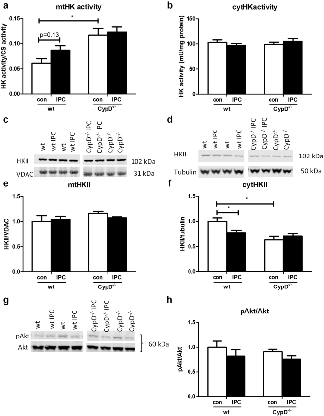
CypD and IPC have no effect on end-ischemic mtHKII in hearts perfused with glucose, pyruvate, lactate and glutamine. (a) End-ischemic mitochondrial HK activity as ratio to CS activity. (b) Cytosolic HK activity normalized to protein content. (c) Representative Western blot images of mitochondrial HKII/VDAC and (d) cytosolic HKII/tubulin. The amount of end-ischemic mitochondrial (e) and cytosolic (f) HKII as ratio of VDAC and tubulin respectively, normalized to WT control. (g) Representative Western blot image and quantification (h) of whole homogenate pAkt/Akt, normalized to WT control. n = 6 per group. Data are shown as mean + SEM. Mann-Whitney U tests were performed to test for significance between 1) control treated WT and CypD−/− animals 2) control and IPC treated animals in WT and 3) CypD−/− animals. *p < 0.05.

