
Figure 1. Distribution of patients according to antibody type.
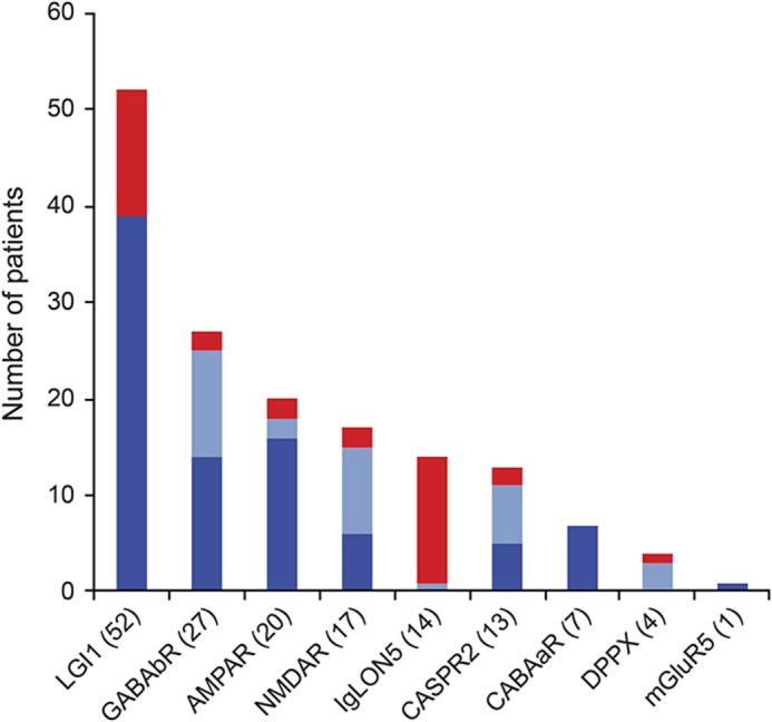
Frequency of patients ≥60 years of age with (blue) or without (red) CSF pleocytosis or inflammatory changes in the brain MRI. Number of cases for each antibody is shown in parentheses. Dark blue in the column indicates number of patients with brain MRI findings compatible with encephalitis; light blue indicates number of cases with normal MRI and CSF pleocytosis.