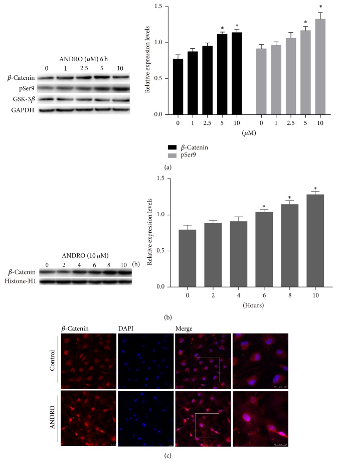
Figure 4.
Activation of the Wnt canonical pathway after treatment with ANDRO. (a) Cells were treated in an ANDRO-containing medium (1, 2.5, 5, and 10 μM) for 6 h followed by 3 days in the neural differentiation medium; the protein expression level of total β-catenin and total GSK-3β and pSer9 detected by Western blotting exhibited a concentration-dependent increase. ∗P < 0.05 compared with the control group. (b) Nuclear protein was extracted at 0, 2, 4, 6, 8, and 10 hours after cells were treated with 10 μM ANDRO, and Western blotting was performed. The protein level of β-catenin in cell nucleus increased with time. ∗P < 0.05 compared with control. (c) Cells were exposed to an ANDRO-containing medium (10 μM) for 6 h followed by 3 days in the neural differentiation medium; immunofluorescence showed that red fluorescence (β-catenin) was superimposed on blue fluorescence (DAPI) in the ANDRO group under a confocal microscope; this was not observed in the control group. Zoomed images of the white square show the details. Immunofluorescence revealed the translocation of β-catenin to the nucleus. Scale bar: 25 μm.

