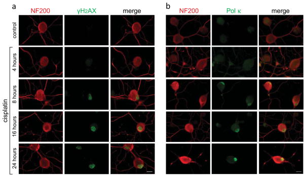
Fig. 3. Temporal patterns of nuclear γH2AX foci formation and DNA Pol κ immunoreactivity induced by cisplatin exposure of DRG neurons.
Gradual increase of nuclear γH2AX fluorescence is observed after 4 h (a, green). Elevated immunoreactivity of Pol κ becomes apparent by 8 h with intensification over the 24-h exposure period (b, green). DRG neurons are identified by immunoreactivity of the high-molecular weight DRG specific neurofilament protein (NF200) in cell bodies and neurites (red). Merged images are shown; scale bar, 10 μm.

