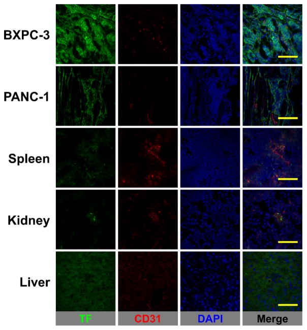
Figure 5.
TF/CD31 immunofluorescence co-staining of pancreatic tumors, spleen, kidney, and liver tissues. TF staining (green channel) was pronounced in BXPC-3 tumors whereas green fluorescence signal was at background levels in PANC-1 tumors, spleen, kidney, and liver. No significant overlap between CD31 (vasculature) and TF staining was observed. Scale bar = 50 μm.

