Figure 1.
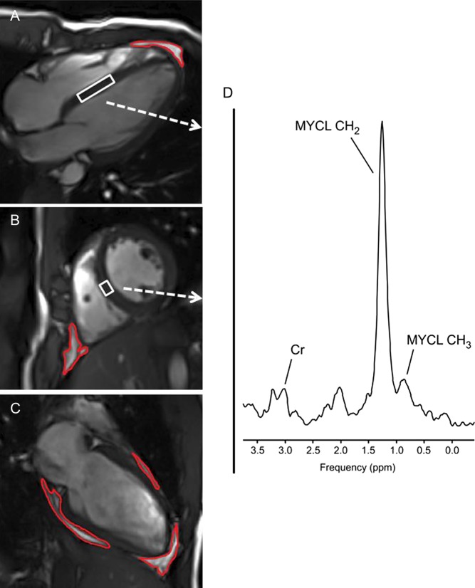
1H MRI and MRS of heart – Cardiac T1-weighted four chambers (A), short axis (B) and two chambers (C) MR images acquired at the end of diastole. Blood and fat tissues show hyperintense (bright). Red contours depict the segmentation of pericardial fat. White boxes within the septum depict the position of volume of interest (VOI) from which 1H MRS (D) is acquired. Spectral lines of methylene (MYCL – CH2) and methyl (MYCL CH3) groups of myocardial lipids as well as the line of creatine (Cr) are annotated.

 This work is licensed under a