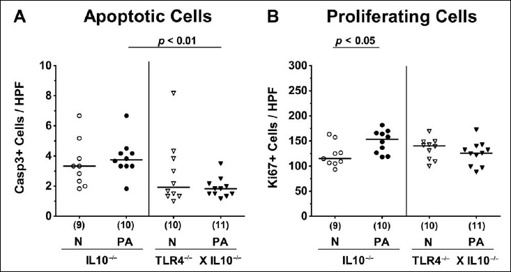
Fig. 3.

Apoptotic and proliferating intestinal epithelial cells following peroral multidrug-resistant P. aeruginosa colonization of secondary abiotic IL10–/– mice lacking TLR4. Secondary abiotic IL10-deficient (IL10–/–; circles) and TLR4-deficient IL10–/– mice (TLR4–/– × IL10–/–; triangles) were perorally challenged with a clinical multidrug-resistant P. aeruginosa strain (PA; closed symbols). Two weeks thereafter, the average numbers of colonic epithelial (A) apoptotic (positive for caspase 3, Casp3+) and (B) proliferating cells (positive for Ki67; Ki67+) were determined in six high power fields (HPF, 400× magnification) per animal in immunohistochemically stained large intestinal paraffin sections. Non-infected mice (N; open symbols) served as negative controls. Numbers of mice (in parentheses), means (black bars), and significance levels (p values) determined by the Mann–Whitney U test are indicated. Data shown were pooled from two independent experiments
