
Figure 6. Comparisons between the scattering best-fit and linear crystal-derived models.
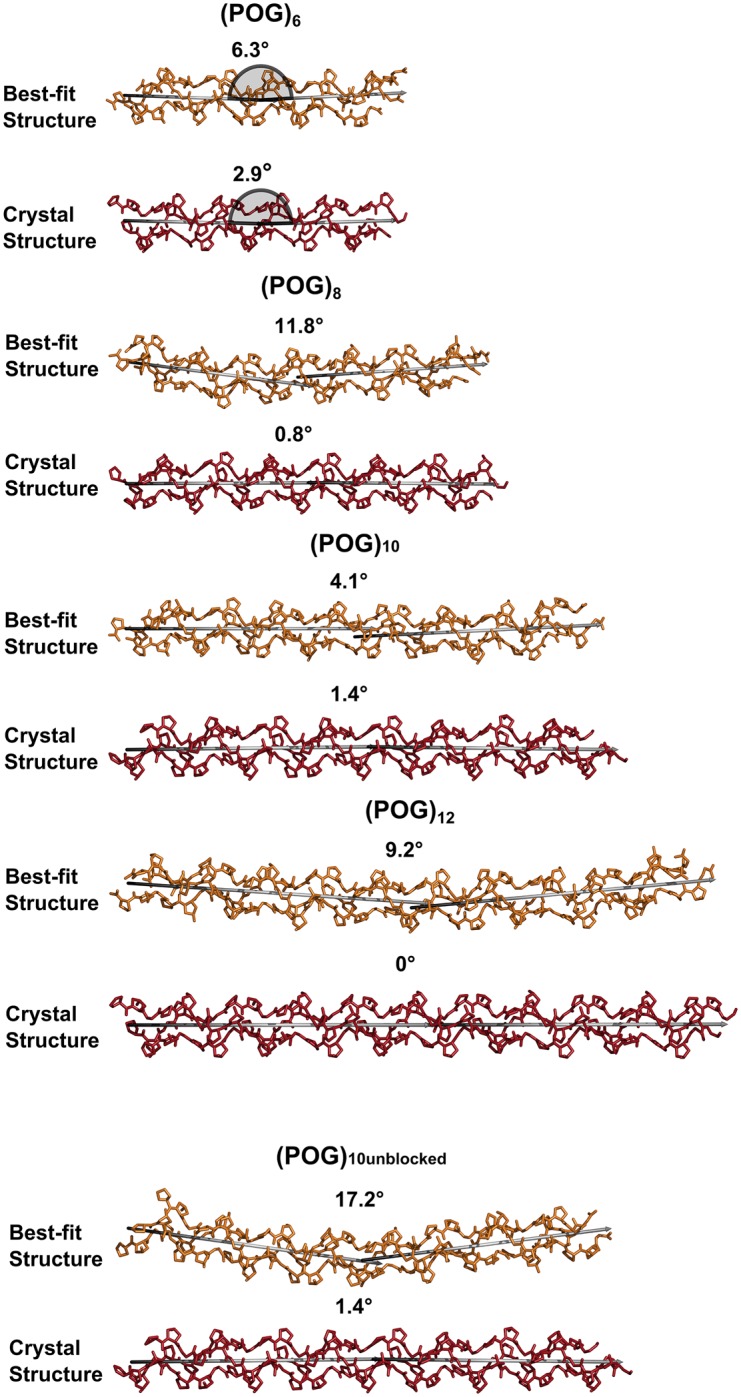
The three polypeptide chains of the best-fit structures for (POG)6-(POG)12 are shown in orange, while those for the linear models are shown in red. In order to monitor the degree of bend in these structures, two verticies shown in grey were fitted to the N-terminal and C-terminal halves of the collagen triple helix, and the angle between the two verticies is displayed above each structure, with 0° representing a linear structure. The grey half circles for (POG)6 define how the angles were measured.