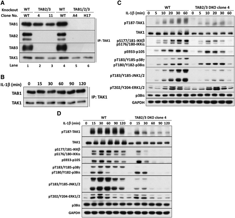
Figure 1. IL-1β signaling in TAB2/3 DKO cells.
(A) Generation of IL-1R* cells lacking TAB2 and TAB3 or all three TAB subunits. TAK1 was immunoprecipitated from the extracts of WT IL-1R* cells (lane 1), two different clones (4 and 11) of cells devoid of TAB2 and TAB3 (lanes 2 and 3) and two different clones (A4 and H17) lacking expression of all three TAB components. Immunoprecipitates were subjected to SDS–PAGE, and immunoblotting as in the Methods with antibodies recognizing TAK1 or each TAB protein. (B) TAB2/3 DKO cells (clone 4 from A) were stimulated with 5 ng/ml IL-1β for the times indicated, and TAK1 immunoprecipitated from the extracts and processed as in A. (C and D) WT or TAB2/3 DKO IL-1R* cells (clone 4 from A) were stimulated with IL-1β for up to 1 h (C) or 2 h (D) as in B, and cell extracts analyzed by SDS–PAGE and immunoblotting with phospho-specific antibodies recognizing phosphorylated (p) serine (pS), threonine (pT) and tyrosine (pY) residues in the activation loops of TAK1, IKKα/β, p105/NF-κB1, JNK1/2, p38α/p38γ and ERK1/ERK2 MAP kinases, or with antibodies recognizing all forms of TAK1, p38α and GAPDH.

