FIGURE 1.
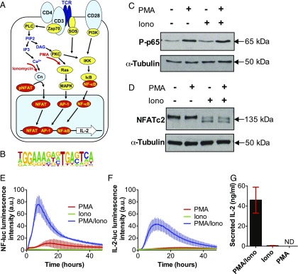
PMA and ionomycin treatment decouple the IKK/MAPK and calcium signaling networks in Jurkat T cells. (A) Schematic representation of TCR signaling network. (B) Composite consensus binding motif for cooperative binding of NFAT and AP-1 (1). (C and D) Immunoblotting analyses of NF-κB phospho-p65 (C) and NFATc2 protein expression levels in Jurkat T cells. Cells stimulated with PMA (20 ng/ml), ionomycin (1 μg/ml), or PMA/ionomycin (20 ng/ml PMA/1 μg/ml ionomycin) for 15 and 60 min, respectively. (E and F) Time series live-cell luminometry analysis of Jurkat T cells expressing reporters of NF-κB–dependent transcriptional activity (E) or IL-2 promoter activity (F) following treatment with PMA, ionomycin, or PMA/ionomycin for 48 h (in triplicates, with error bars indicating SDs). (G) Concentration of IL-2, measured by ELISA, secreted by Jurkat T cells treated with PMA/ionomycin (20 ng/ml PMA/1 μg/ml ionomycin), ionomycin (1 μg/ml), or PMA (20 ng/ml). Means (±SDs) of triplicate experiment is shown in red. ND, not detected.

