FIGURE 1.
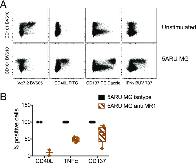
Human MAIT cell activation leads to MR1-dependent CD40L upregulation. Whole blood was incubated for 16 h with 5-A-RU and MG in the presence of protein transport inhibitors. (A) Expression of Vα7.2 TCR, CD40L, CD137, and IFN-γ was detected by intracellular staining and is depicted in the FACS dot plots, in parallel with CD161 expression. (B) Effect of MR1 blockade on CD40L (n = 2), TNF-α (n = 5), and CD137 (n = 5) expression in the intracellular assay performed as depicted in (A). Data are mean ± SD. For CD137-expressing cells, the ratio of mean fluorescence intensity with and without anti-MR1 is plotted, because the overall percentage of positive cells remains unchanged (see also Supplemental Fig. 4B). Statistical analysis was not performed for CD40L because of the small sample size. p = 0.06 for TNF-α and CD137, Wilcoxon signed-rank test.

