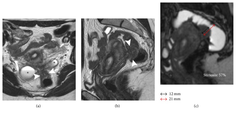
Figure 1.
32-year-old patient with DPE and infiltrative nodule requiring segmental resection. (a) Axial T2W image, (b) sagittal T2W image, and (c) MR-Colonography. Pelvic tethering involving the ovaries and the rectum with Douglas pouch obliteration. An infiltrative nodule (short axis 12 mm) is visible on the anterior wall of the rectum (arrowheads). Presence of bilateral endometriomas (∗). MR-Colonography demonstrates a stenosis of 57% (c).

