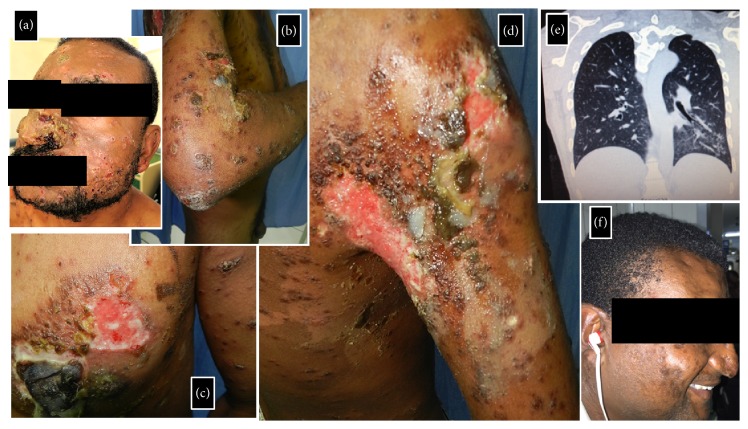
Figure 1.
Clinical images of our 34-year-old male patient who presented with a 2-month history of rapidly spreading multiple cutaneous lesions. Annular brownish papules and plaques, with or without a scale crust, scattered over the face (a), chest (c/d), and extremities (b/d). Some larger lesions assumed a rupioid aspect (c). Large reddish shallow ulcers in right mammary (c) and right anterior axillary (d) regions. Some lesions drained a seropurulent discharge. Ground grass opacities are seen over the superior segment of the left inferior lobe (e). Complete remission of cutaneous lesions after treatment (f).

