Figure 3.
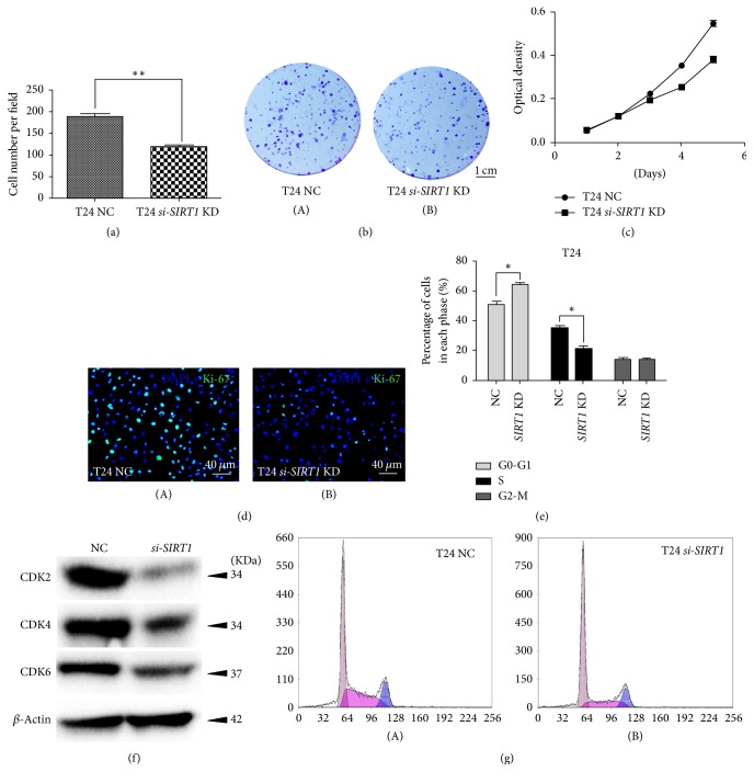
Downregulation of SIRT1 repressed BCa cell proliferation and induced cell cycle arrest. (a) Clone number in each well was counted and statistically analyzed in the clonogenic survival assay. ∗∗p < 0.01. (b) Clonogenic survival assay revealed cell survival of BCa cells after treatment of SIRT1-target-specific-siRNA (SIRT1 KD) and control-siRNA (NC), cultured in 6-well plates for 14 days. (c) MTT assay was used to measure the viability of BCa cells treated by SIRT1-target-specific-siRNA (SIRT1 KD, line linking squares) and negative-control-siRNA (NC, line linking circles). All shown values were mean ± SD of three measurements and repeated three times with similar results, ∗p < 0.05. (d) Cell proliferation of BCa cells treated by SIRT1-target-specific-siRNA (B) and negative-control-siRNA (A) was assayed by Ki-67 staining (green). Nuclei (blue) were stained by DAPI. (e) Statistical analysis of percentages (%) of BCa cell populations at different stages of cell cycles. All shown values were mean ± SD of three measurements and repeated three times with similar results. ∗p < 0.05. (f) Western blot analysis of proteins involved in G0-G1 cell cycle regulation (CDK2, CDK4, and CDK6) in the BCa cells. β-Actin abundance was used as a control. (g) Flow cytometry analysis result for BCa cells treated with negative-control-siRNA (A) and SIRT1-target-specific-siRNA (B) for 48 h. The scale bar for (b) is 1 cm and for (d) is 40 μm.

