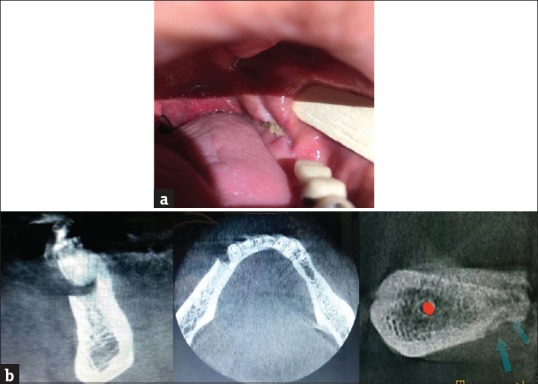
Figure 1.

(a) Osteonecrosis of the jaw. (b) Osteonecrosis of the jaw in cone beam computed tomography, cone beam computed tomography cross section related to posterior part of mandibular ridge in the left side showed the necrotic bone which got separated from normal bone
