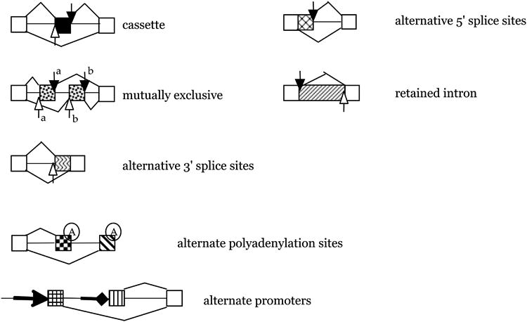
Figure 1. Alternative splicing modes.

Constitutive exons are shown as white boxes, introns as vertical lines. Open arrows indicate the 3′ splice site, closed arrows the 5′ splice site. A black box indicates a cassette exon, the most frequent form of alternative splicing. Hatched boxes indicate other splicing modes. Alternative polyadenylation sites and alternate promoters are shown as two other mechanisms to increase mRNA diversity. They are mechanistically different from alternative splicing.
