Figure 1.
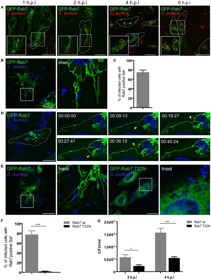
S. aureus induces formation of tubular structures decorated with Rab7. (A) GFP-Rab7wt stably transfected CHO cells were infected with S. aureus wt for 1 h (1 h.p.i) and then washed and incubated for additional 1 h (2 h.p.i), 3 h (4 h.p.i), or 5 h (6 h.p.i) to allow bacteria replication. Bacteria were labeled with rhodamine-red (B) GFP-Rab7 CHO cells were infected for 30 min with S. aureus and living cells were imaged. (C) Quantification of the percentage of infected cells that presented Saf formation. (D) Movie showing CHO cells overexpressing GFP-Rab7 and infected with S. aureus. Yellow arrow heads indicate Saf extension, branching and phagosome contact. Relative time-points are expressed as h:min:s. (E) CHO GFP-Rab7wt and CHO GFP-Rab7T22N (dominant negative mutant) stably transfected cells were infected for 1 h with S. aureus wt. Bacteria were labeled with Hoechst. (F) Quantification showing the percentage of cells with Rab7 positive Saf in cells overexpressing Rab7wt or Rab7 T22N. (G) The graph shows the quantification of the number of CFU/ml in CHO cells stably transfected with GFP-Rab7 (black bars) or GFP-Rab7T22N (gray bars) and infected with S. aureus wt for 2 or 4 h. In all cases data show mean ± SEM from three independent experiments (n = 150 cells/condition). *P ≤ 0.01, ***P ≤ 0.0001 from two-tailed Student t-test. Scale bar: 20 μm.

