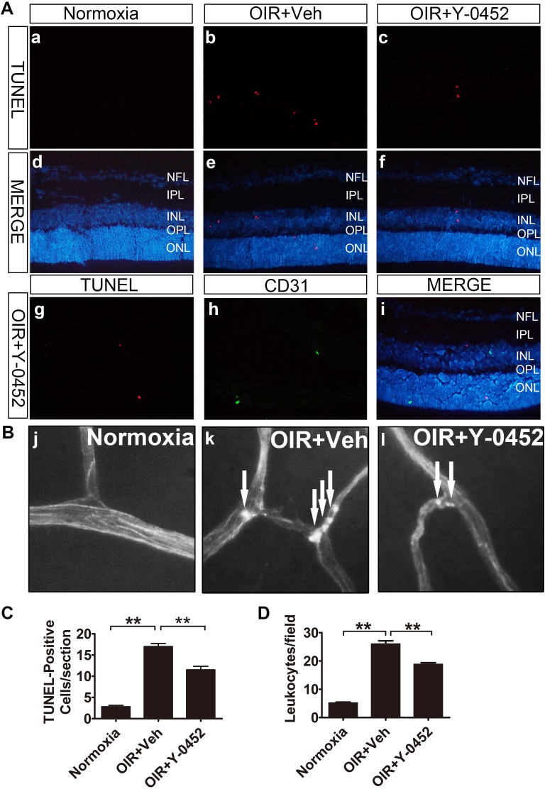
Figure 8.
Effects of Y-0452 on retinal apoptosis and inflammation in OIR mice. WT OIR mice were intraperitoneally injected with Y-0452 (10 mg/kg/d) from P12 to P16 or the same volume of vehicle. (A) Representative images of retinal apoptotic cells in OIR mice (normoxia: [a, d], OIR+Veh: [b, e], OIR+Y-0452: [c, f], costaining with anti-CD31: [g–i]). Retinal apoptotic cells were labeled by TUNEL (red), and nuclei stained with DAPI (blue). CD31 (green) cells were co-immunostained with TUNEL (red) and DAPI (blue) in OIR retinal sections. (B) Representative images of retinal adherent leukocytes in OIR mice (white arrows indicate adherent leukocytes). (C) Quantification of retinal TUNEL-positive cells in OIR mice from retinal sections (n = 6; *P < 0.05, **P < 0.01 versus vehicle, 1-way ANOVA). (D) Quantification of retinal adherent leukocytes in OIR mice was performed from ×40 magnification images. Adherent leukocytes were counted in six random fields per retina (n = 6; *P < 0.05, **P < 0.01 versus vehicle, 2-way ANOVA).

