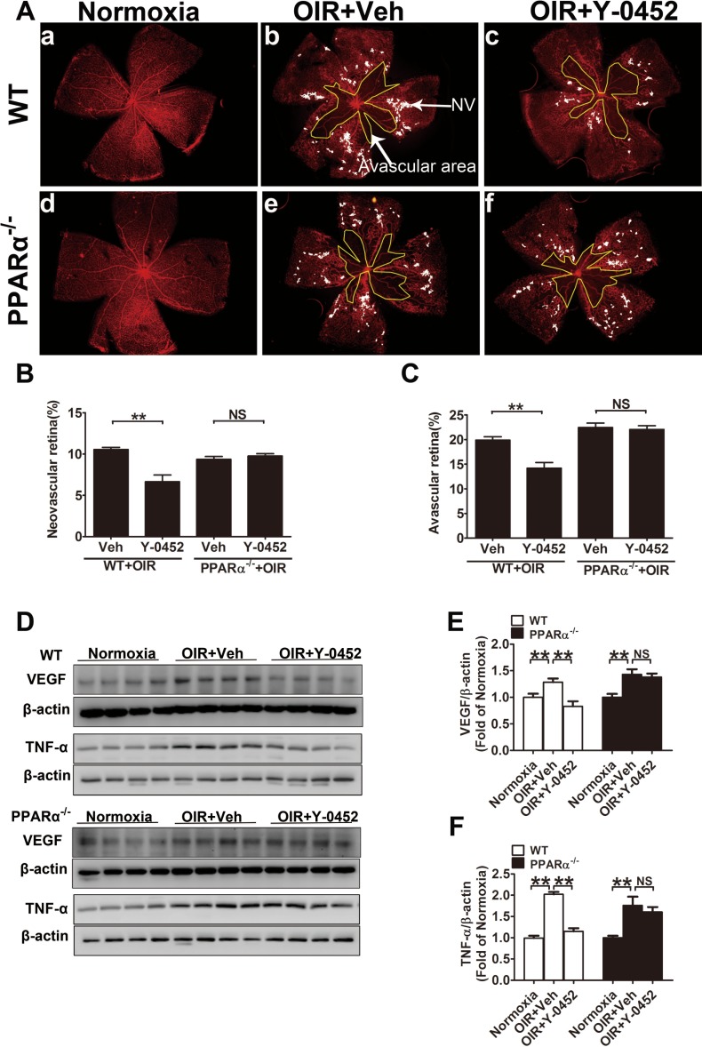
Figure 9.
Effect of Y-0452 on retinal neovascularization in OIR mice. WT and PPARα−/− OIR mice were intraperitoneally injected with Y-0452 (10 mg/kg/d) from P12 to P16 or the same volume of vehicle. The retinas were fixed and stained with Isolectin B4. Areas of retinal neovascularization and vaso-obliteration were quantified under a fluorescence microscope. (A) Representative images of retinal neovascularization and avascular areas in WT (a–c) and PPARα−/− OIR mice (d–f). The white dot–marked area indicates neovascular retina and the yellow line–marked area indicates avascular area. (B, C) Quantification of retinal neovascularization and avascular areas. (D) Retinal VEGF and TNF-α levels were measured by Western blot analysis. (E, F) Quantification of VEGF levels in WT and PPARα−/− OIR mice (n = 8; *P < 0.05, **P < 0.01, 2-way ANOVA).

