Figure 3.
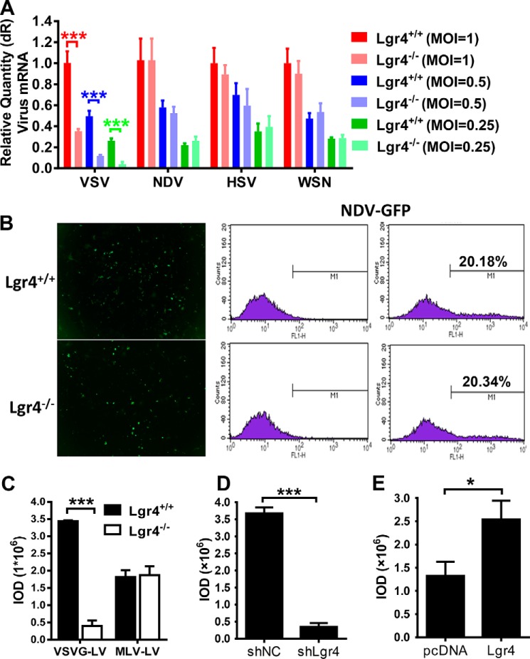
Lgr4-mediated virus infection is dependent on VSV-G. A, Q-PCR analysis of indicated virus in Lgr4+/+ and Lgr4−/− macrophages infected with VSV, NDV, HSV-1, or WSN at an m.o.i. of 1, 0.5, and 0.25 for 8 h. B, quantitative flow cytometry analysis of Lgr4+/+ and Lgr4−/− MEFs infected with NDV-GFP (m.o.i. = 10) for 24 h. C–E, Lgr4+/+ and Lgr4−/− MEFs were infected with VSV-G protein-coated lentivirus or MLV protein-coated lentivirus expressing GFP for 48 h (C). Lgr4 control and knockdown RAW 264.7 cells were infected with VSV-G protein-coated lentivirus expressing GFP for 48 h (D). Control and Lgr4 overexpressing RAW 264.7 cells were infected with VSV-G protein-coated lentivirus expressing GFP for 48 h (E). Digital images were captured using an Olympus IX71 inverted fluorescence microscope with DP2-BSW imaging software. Results are shown as the mean ± S.D. (n = 3). The mean fluorescence density was determined by IOD. Data are representative of at least three independent experiments. Asterisks indicate statistical significance: ***, p < 0.001; *, p < 0.05.

