Figure 4.
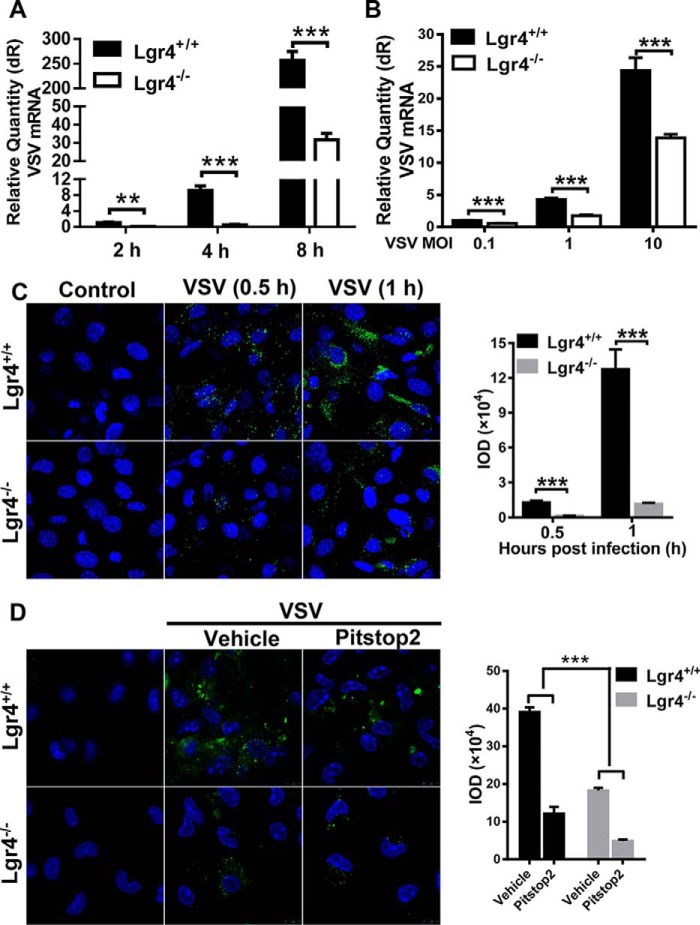
VSV entry is decreased in Lgr4-deficient cells. A, Q-PCR analysis of VSV RNA in Lgr4+/+ and Lgr4−/− peritoneal macrophages at the indicated times (m.o.i. = 1). B, Q-PCR analysis of VSV RNA in Lgr4+/+ and Lgr4−/− peritoneal macrophages infected with VSV for 1 h at 4 °C at the indicated m.o.i. C, immunofluorescence staining of VSV (green) on the surface of Lgr4+/+ and Lgr4−/− MEF cells infected by VSV (m.o.i. = 1000) for 30 min or 1 h at 4 °C. D, immunofluorescence staining of VSV (green) on the surface of Lgr4+/+ and Lgr4−/− MEF cells infected by VSV (m.o.i. = 1000) for 6 h after treatment with pitstop2 (30 μm) 10 min at 37 °C. DAPI, blue. Data are representative of at least three independent experiments. Asterisks indicate statistical significance: ***, p < 0.001; **, p < 0.01.

