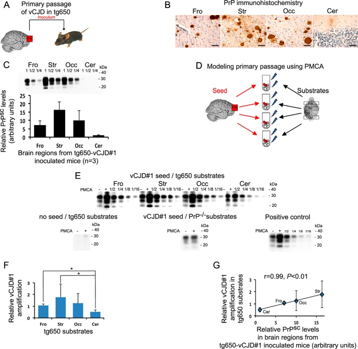
Figure 1.
rsPMCA modeling of region-specific targeting was observed after primary passage of vCJD to transgenic mice expressing human PrPC. A, schematic representation of in vivo experiment. B and C, PrPSc accumulation in different brain regions of vCJD-inoculated tg650 mice that overexpress M129 human PrPC as illustrated by PrP immunochemistry (B) and revealed by Western blotting (C). D, schematic representation of in vitro modeling. E, Western blot illustration of the amplification obtained using vCJD seed and brain substrates from tg650 mice. No amplification was observed when the vCJD seed was omitted and when frontal substrates from PrP knock-out mice were used as substrate. F, relative rsPMCA amplification obtained with vCJD and the different substrates from tg650 mice. G, relative rsPMCA amplification plotted as a function of relative PrPSc level measured in the corresponding brain areas of vCJD-inoculated tg650 mice. Scale bar, 50 μm. Fro, frontal cortex; Str, striatum; Occ, occipital cortex; Cer, cerebellar cortex; PMCA−, no PMCA was applied; PMCA+, one round of PMCA was applied; 1/2, 1/4, 1/8, 1/16, rsPMCA product dilution. Error bars represent S.D.; *, p < 0.05. PMCA results are representative of four independent experiments performed in duplicates.

