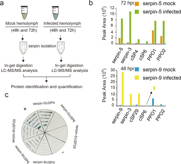
Fig 5. Proteomics analysis of serpin-associated proteins.
(a) Schematic for isolation of serpin-associated proteins from cell-free hemolymph. (b) Proteins eluted from immunoaffinity columns were identified by in-gel trypsin digestion and LC-MS/MS analysis. The arrow point to magnified image of cSP6 peak area. Floating yellow bar indicate the magnified peak area of cSP6. (c) Yeast-two-hybrid assay validates the binding between the identified serpins and cSPs in SD-Leu-Trp-His medium with X-α-gal. +: positive control, -: negative control.

