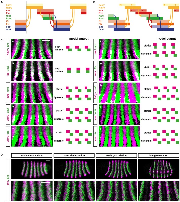
Fig 4. Dynamic patterning of the primary pair-rule genes.
(A) Regulatory schematic showing the predicted phasing of the primary pair-rule stripes during cellularisation, assuming static, partially overlapping domains of Hairy and Eve. Pale colours represent transcript domains; intense colours represent protein domains; hammerhead arrows represent repressive interactions; grey vertical lines indicate the span of a double-segment pattern repeat. Cross-regulatory interactions are from the early network (compare Fig 1A, left). For simulation output, see S1 Movie. (B) Regulatory schematic showing the predicted phasing of the pair-rule stripes during cellularisation, assuming dynamic, partially overlapping domains of Hairy and Eve. hairy and eve domains shift anteriorly over time, resulting in offsets between transcript and protein domains. Colours, etc., as for (A). For simulation output, see S2 Movie. (C) Comparisons between real and predicted phasings of the primary pair-rule stripes. Double FISH images show lateral views of stripes 2–6 (anterior left, dorsal top) in mid-cellularisation stage embryos. In the bottom half of each image, the 2 channels have been thresholded, making regions of overlap easier to see. Scale = 50 μm. Diagrams to the right of each image show the stripe phasings predicted by static (top) or shifting (bottom) gap inputs, respectively (compare A and B). For panels (A) and (B), the 2 models predict the same relative pattern. In all other panels, the models predict different relative patterns. (D) Simultaneous visualisation of eve transcript (magenta) and Eve protein (green) in embryos at 4 different stages. Rightmost panels show a ventrolateral view; all other panels show lateral views. Upper panels show whole embryo views; lower panels show enlarged views of stripes 2–6. In stripes 3 onwards, the protein domains lag behind the transcript domains until late gastrulation, indicating that the anterior boundaries of the eve stripes shift anteriorly until early gastrulation. In stripe 2, the anterior boundary stabilises significantly earlier. Scale = 50 μm. Abbreviations: eve, even-skipped; FISH, fluorescent in situ hybridization.

