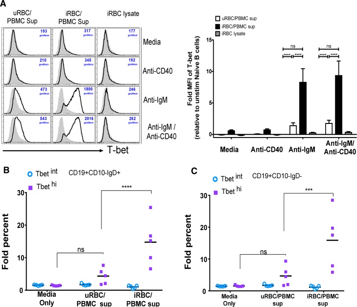
Fig 10. Supernatants of PBMCs stimulated with P. falciparum-infected RBCs plus BCR cross-linking drive T-bet expression in B cells.
(A-C) PBMCs of healthy U.S. adults (n = 5) were stimulated in vitro with P. falciparum-infected red blood cell (iRBC) lysate or uninfected red blood cell (uRBC) lysate for 3 days. The resulting supernatants or the iRBC lysate alone were transferred to PBMCs from the same U.S. adults (n = 5) in the presence of media alone, anti-IgM, anti-CD40, or both, followed by staining for T-bet, CD10, CD19 and IgD. (A) Fold change in T-bet MFI in stimulated naïve B cells relative to unstimulated naïve B cells (left, representative histograms). Fold change in percentage of T-bet intermediate (T-betint) and T-bet high (T-bethi) (B) naïve B cells and (C) memory B cells after BCR cross-linking with anti-IgM/G/A in the presence of media alone, uRBC/PBMC supernatant or iRBC/PBMC supernatant, relative to unstimulated cells. Horizontal bars and whiskers represent means or median and SE. p values were determined by paired Student’s t test with Bonferroni adjustments where appropriate. ****P<0.0001, ***P<0.001, **P<0.01, *P<0.05, ns = not significant.

