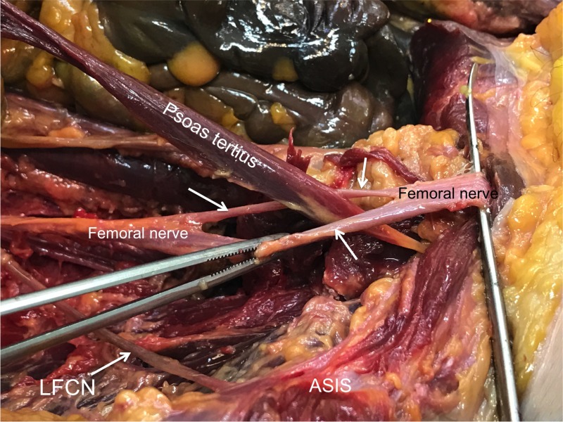
Figure 1. Right posterior abdominal wall of a 74-year-old female cadaver.
The psoas tertius muscle has been disconnected from its origin and is shown traveling distally through the femoral nerve (white label indicates proximal nerve and the black label indicates distal nerve prior to traveling deep to the inguinal ligament) to fuse beyond this point with the tendon of iliopsoas. The perforation of the femoral nerve is shown with arrows. For reference, the right lateral femoral cutaneous nerve (LFCN) is shown traveling over iliacus toward the anterior superior iliac spine (ASIS).

