FIGURE 6.

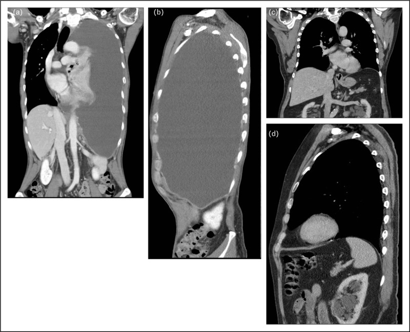
(a, b) CT scan (coronal and sagittal views) shows a large pleural effusion pushing down and inverting the left diaphragm. (c, d) The left diaphragm reverts to its normal shape and position following complete evacuation of the effusion.

(a, b) CT scan (coronal and sagittal views) shows a large pleural effusion pushing down and inverting the left diaphragm. (c, d) The left diaphragm reverts to its normal shape and position following complete evacuation of the effusion.