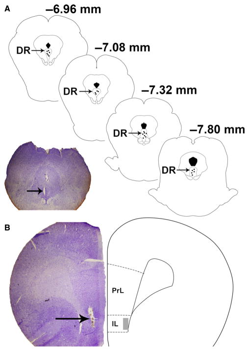
Fig. 1.
(A) Diagram illustrating the location of recording electrodes in the dorsal raphe (DR) (ML 0 mm, AP +12 mm, and DV +7–8 mm relative to bregma in the 32° plane). The section of the brain on the left (40 μm) has been stained with cresyl violet and illustrates the recording electrode lesion (arrow). (B) Illustration (right) of the stimulating electrode location in the infralimbic region (IL) of the medial prefrontal cortex (mPFC) along with cresyl violet histological section (left) of the same region with electrode lesion (arrow).

