
Fig. 2.
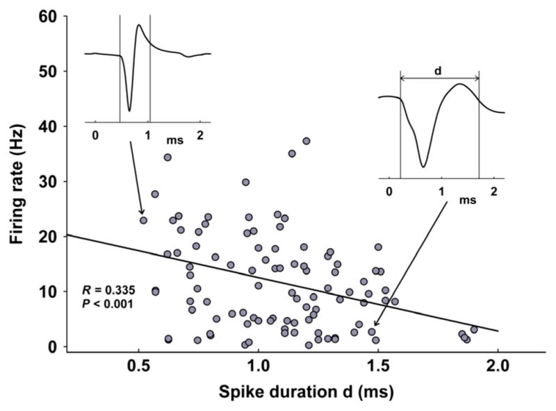
All recorded neurons in the dorsal raphe plotted based on spike duration (d in waveform inset, x-axis, in ms) and baseline firing rate (y-axis, Hz). The two insets are examples of a slow-firing (right) and fast-firing neuron (left). The distribution of frequencies revealed a significant correlation between cellular firing rate and spike width, where slower firing cells tended to have broader spikes, while fast spiking neurons were found to have shorter spike width (R = 0.330, P < 0.001, n = 102, 35 rats).