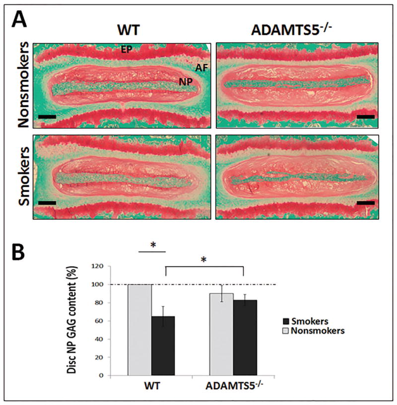
Figure 4. Depletion of ADAMTS5 reduced disc proteoglycan loss in a mouse model of tobacco smoking.

(A) Representative images of Safranin O/fast green stained discs (bar=250 μm) of WT and ADAMTS5-deficient mice treated with or without tobacco smoke for six months. Red stain intensity indicates the level of proteoglycan. Nucleus pulposus (NP), annulus fibrosis (AF), and cartilaginous endplate (EP) regions are indicated. (B) DMMB quantitative assay for total GAG content from NP tissue of smoke-exposed (smokers) WT and ADAMTS5-deficient mice and their respective untreated controls (nonsmokers). Average values ± stdev of eight exposed mice and eight unexposed controls are shown. * denotes p<0.05.
