
Figure 6.
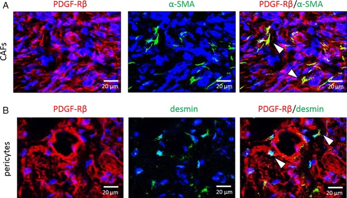
Fluorescence double‐labeled immunohistochemistry (IHC) of Caki‐1 human kidney cancer cells grown in nude mice. Representative images show IHC for α‐SMA (CAFs marker, Fig. 6A), desmin (pericyte marker, Fig. 6B) in green, and PDGFR‐β in red. Expression of PDGFR‐β in CAFs or pericytes is indicated by an arrow‐head. Scale bars: 20 μm (A, B).