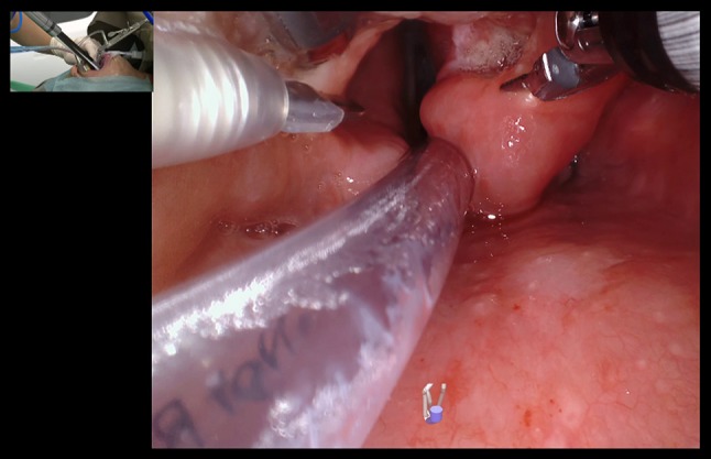
Fig. 2.

da Vinci SP in use for examination of the supraglottis through an internal view. The monopolar spatula tip is seen on the left, the Maryland bipolars are opened at the right aryepiglottic fold exposing the tumor, while the fenestrated bipolar is retracting the epiglottis superiorly. The insetted figure shows the assistant holding a Yankaur suction and the location of the port about 10 cm from the oral opening
