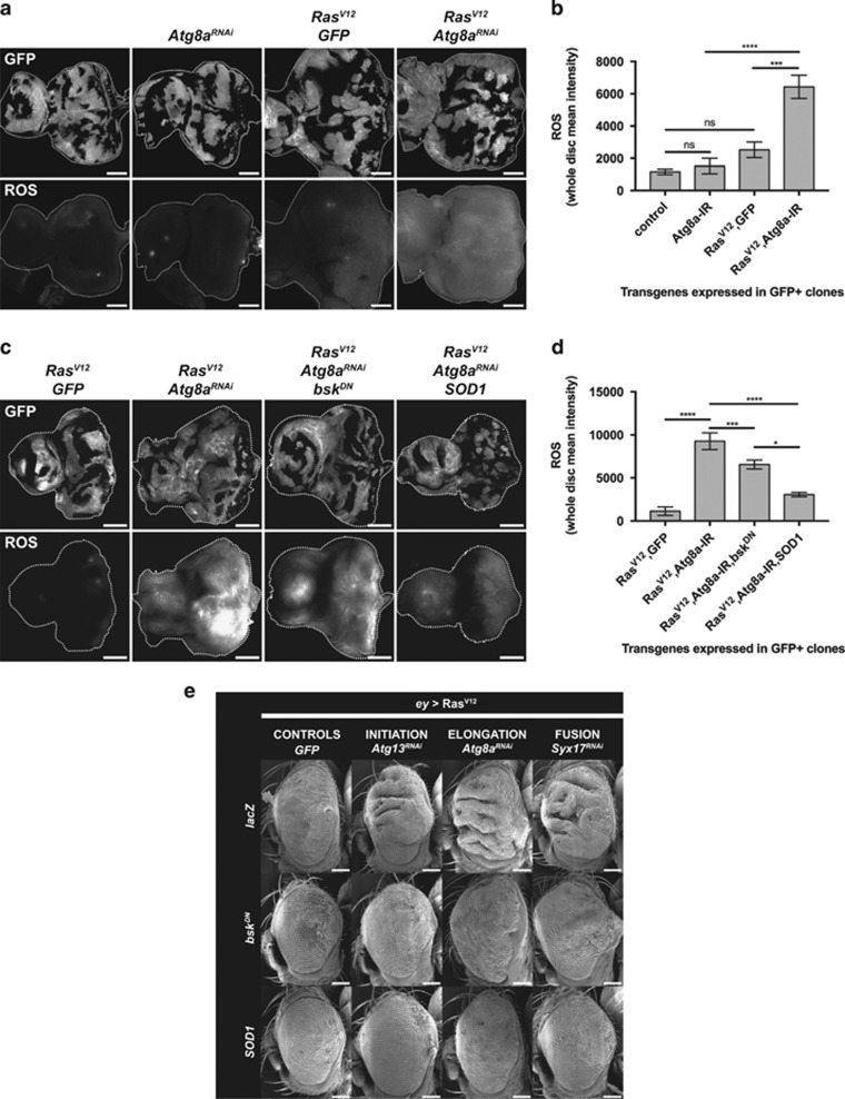
Figure 8.
Upon autophagy inhibition, ROS accumulate in Ras-activated tissue and lead to cooperative tissue overgrowth via JNK activation. (a) Oxidative stress levels in eye-antennal mosaic discs as detected by CellROX Deep Red staining. Autophagy inhibition using Atg8aRNAi did not augment ROS levels compared with control discs. Ras activation (RasV12 GFP) slightly increased ROS levels. However, massive accumulation of ROS was detected in RasV12Atg8aRNAi mosaic tissue. Interestingly, ROS are not limited to RasV12Atg8aRNAi clones, but spread in the whole disc, suggesting a non-cell-autonomous regulation of oxidative stress levels in RasV12AtgRNAi mosaic tissue. Scale bar: 50 μm. (b) Quantification of ROS levels in whole discs from (a). (c) ROS levels in RasV12 Atg8aRNAi mosaic discs are reduced upon SOD1 overexpression, but not upon JNK blockage, confirming that JNK activation is downstream of ROS accumulation in RasV12 Atg8aRNAi discs. Note that overexpression of SOD1 rescues both cell-autonomous and non-cell-autonomous accumulation of ROS, and restores the size and shape of RasV12 Atg8aRNAi eye discs. (d) Quantification of ROS levels in whole discs from (c). (e) Scanning electron micrographs of adult eyes expressing RasV12 and GFP (first column), Atg13RNAi (second column), Atg8aRNAi (third column) or Syx17RNAi (fourth column), in combination with control lacZ (first row), bskDN (second row) or SOD1 (third row). RasV12AtgRNAi eye overgrowth is rescued by overexpression of bskDN or SOD1, showing that autophagy-impaired RasV12-driven tumourigenesis is due to elevated ROS levels and JNK signalling. Note that overexpression of SOD1 and bskDN also rescues Ras overgrowth alone (first column). Error bars=s.e.m. Statistics: one-way ANOVA with Tukey's multiple correction.

