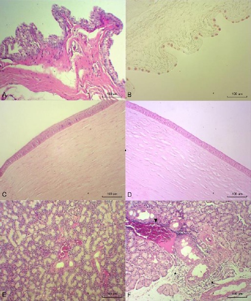
Fig. 6.

Photomicrographs of the conjunctiva, cornea and lacrimal gland at T12. (A) LO group (OD): conjunctiva with large numbers of goblet cells (hematoxylin-eosin (HE), 100x). (B) LO group (OS): goblet cells (PAS-alcian blue, 100x). (C) LO group (OD): normal cornea (HE, 100x). (D) LO group (OD): cornea with edema (HE, 100x). (E) FO group (OD): normal lacrimal gland (HE, 100x). (F) FLO group (OS): lacrimal gland with inflammatory infiltration (arrows) and vascular congestion (arrowhead) (HE, 100x).
