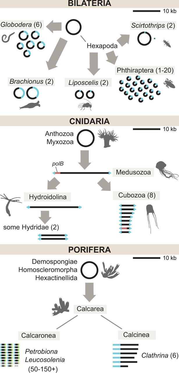
Fig. 2.—

Evolution of linear and multipartite mitochondrial DNA in animals. Gray arrows indicate independent reorganization events in the common ancestor of the indicated group. Blue segments indicate regions of homology shared across chromosomes. Numbers in parentheses indicate the range of chromosome numbers (greater than 1) found in the group. Top: Multipartite mtDNA in Bilateria. Not shown: An unknown number of mini-circle chromosomes in Dicyema (Watanabe et al. 1999). Middle: Linear and multipartite mtDNA in Cnidaria. The red segment denotes the presence of a polB coding sequence in some Medusozoa. Arrowheads at the ends of linear chromosomes signify inverted terminal repeat sequences. Bottom: Multipartite linear mtDNA in Porifera. Faded chromosomes and ellipses indicate the inferred presence of an uncertain number of additional chromosomes. Blue and green segments in Leucosolenia and Petrobiona show distinct end sequences shared across chromosomes, but which are not homologous between the two genera. Genomes are drawn to scale with respect to the 10 kb scale bar in the upper right part of each panel.
