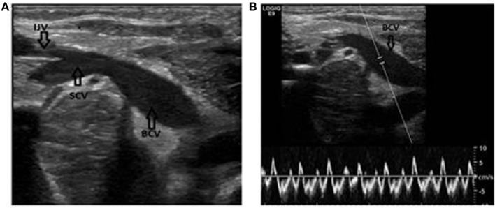
Figure 2.
Identification of the subclavian and brachiocephalic veins. (A) Long axis view of the veins. Notice the confluence of the brachiocephalic vein and the internal jugular vein. IJV, internal jugular vein; SCV, subclavian vein; BCV, brachiocephalic vein. (B) Same patient. The Doppler is used to verify the flow in the vein. Notice how the flow changes with the respiration.

