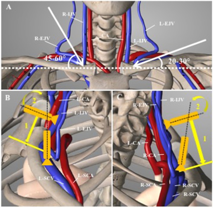
Figure 3.
Anatomical view of the cervicothoracic region. (A) Frontal view outlining the different angles of puncture between right and left sub-clavian (SCV) and brachiocephalic (BCV) veins. CA, carotid artery; IJV, internal jugular vein; EJV, external jugular vein. (B) Left SCV approach: the probe is slided (1) down perpendicular to the IJV and tilted anteriorly (2) toward the L-SCV. Noted that the left subclavian artery (L-SCA) is running posteriorly to the aorta. (C) Right SCV approach: Similarly, to the left side approach, the probe is slided down the IJV (1), than tilted anteriorly (2). Noted the close relation of the right-SCV and right-SCA (adapted from Essential Anatomy, V5.0.3, 3D4Medical.com).

