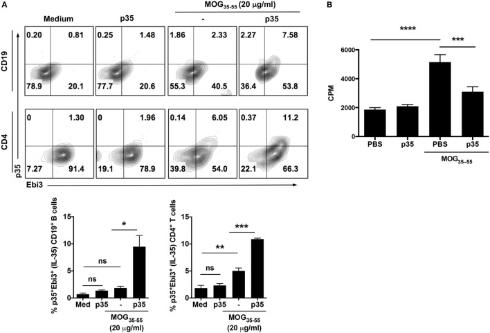
Figure 2.
IL-12p35 induces expansion of IL-35-expressing B cells in the spleen during experimental autoimmune encephalomyelitis (EAE). (A) EAE was induced in mice by active immunization with MOG35–55-peptide in complete Freund’s adjuvant, and the mice were treated with IL-12p35 or PBS. Spleen cells isolated from the mice on day 17 after EAE induction were restimulated ex vivo with MOG35–55 in presence or absence of IL-12p35 for 72 h and were analyzed by intracellular cytokine staining analysis (A) or lymphocyte proliferation assay (B). (A) For intracellular cytokine assay, cells were gated on CD19+ or CD4+ cells, and numbers in the quadrants indicate the percentages of T or B cells expressing IL-35 (p35 and Ebi3). The data are presented as the mean ± SEM of three determinations. ****p < 0.0001; ***p < 0.001; **p < 0.01; *p < 0.05, significantly different from PBS-treated. (B) Proliferative responses assessed by 3H-thymidine incorporation assay were analyzed in five replicate cultures, and data presented as mean value of CPM of the five replicate cultures. Results represent at least three independent experiments. *p < 0.05, **p < 0.01, ***p < 0.001, ****p < 0.0001 [Student’s t-test (two-tailed)].

