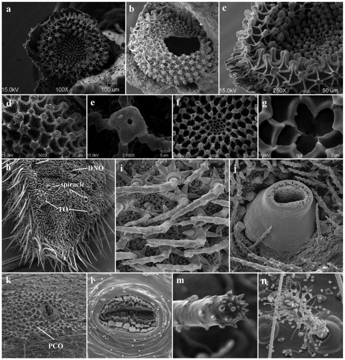
Fig. 3.
SEMs of the P. ripartii egg (a–g), larva (h–j), and pupa (k–n). (a) general shape and structure of the freshly laid egg; (b) egg shell after hatching; (c) concentric zones with three different types of reticulation; d, lateral zone with stalks and low ribs; (e) aeropyle; (f) transitional zone; (g) micropylar pit; (h) terminal segments of the last instar (DNO, TO); (i) seta with stellate base; (j) raised spiracle with semielliptical opening; (k) spiracle surrounded by PCO; (l) lamellate, branched papillae of spiracle; (m) cremaster seta with apical, blunt spine-like projections; (n) dendritic seta on the fifth abdominal segment.

