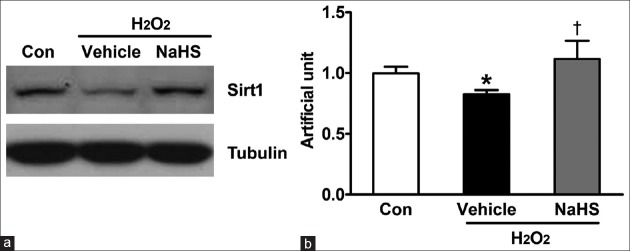
Figure 3.
NaHS induces Sirt1 expression in neonatal mouse cardiomyocytes. Neonatal mouse cardiomyocytes were plated in six-well plates. After 24 h, cells were pretreated in free-serum medium with or without 100 μmol/L NaHS for 12 h. Consequently, cells were washed twice with PBS and were recovered for 24h by changing into FBS-MEM, followed by exposure to free-serum medium without or with 600 μmol/L H2O2 for another 4 h. (a) Cells were lysed and Sirt1 protein expression level is determined using Western blot analysis. (b) The bar charts indicating the different intensities of Sirt1 between groups. Values were normalized against the control values. The data shown are mean ± SE of three independent experiments (*P < 0.05 vs. Con, †P < 0.01 vs. vehicle). PBS: Phosphate-buffered saline; FBS: Fetal bovine serum; MEM: Modified Eagle's medium; SE: Standard error; Con: Control; NaHS: Sodium hydrosulfide; Sirt1: Sirtuin 1.

