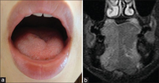
Figure 2.

Case of hypoglossal nerve palsy after surgery, (a) photograph taken early after surgery demonstrating the hypoglossal nerve palsy on the right side. (b) Short-tau inversion recovery of magnetic resonance imaging late after surgery demonstrating high signal intensity on the right side of the tongue, suggesting hypoglossal nerve palsy
