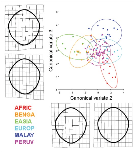
Figure 13.

Results of a canonical variate analysis among 121 dry adult human crania separated into six populations (AFRIC: n = 8; BENGA: n = 15; EASIA: n = 7; EUROP: n = 17; MALAY: n = 15; PERUV: n = 59) with 90% confidence ellipses corresponding to each respective population. The second and third canonical variates, illustrating the variation among groups (scaled by the inverse of the within-group variation), account for 41.87% of the total variance (canonical variate 2: 22.63% of total variance; canonical variate 3: 19.24% of total variance). Thin plate splines along the canonical variate axes illustrate shape change between the populations. Canonical variate 2 is characterized by a unilateral inverse curve change between the anterior right and posterior right aspects of the foramen. Canonical variate 3 is mainly characterized by an inverse relationship between the posterior left and posterior right aspects of the foramen. With regard to the separation of crania among the different populations, the crania of African-descent (AFRIC) are completely separate from the, East Asian (EASIA), Peruvian (PERUV), and European-descent (EUROP) populations. Likewise, there is minimal overlap between the African-descent and Bengali populations. The foramen magnum of the Pervuian population was also completely separated from those of the Malayan population. The Bengali population had some overlap with all other populations. Foramina located near the centroids of the confidence ellipses are presented to illustrate representative samples from each of the six populations
