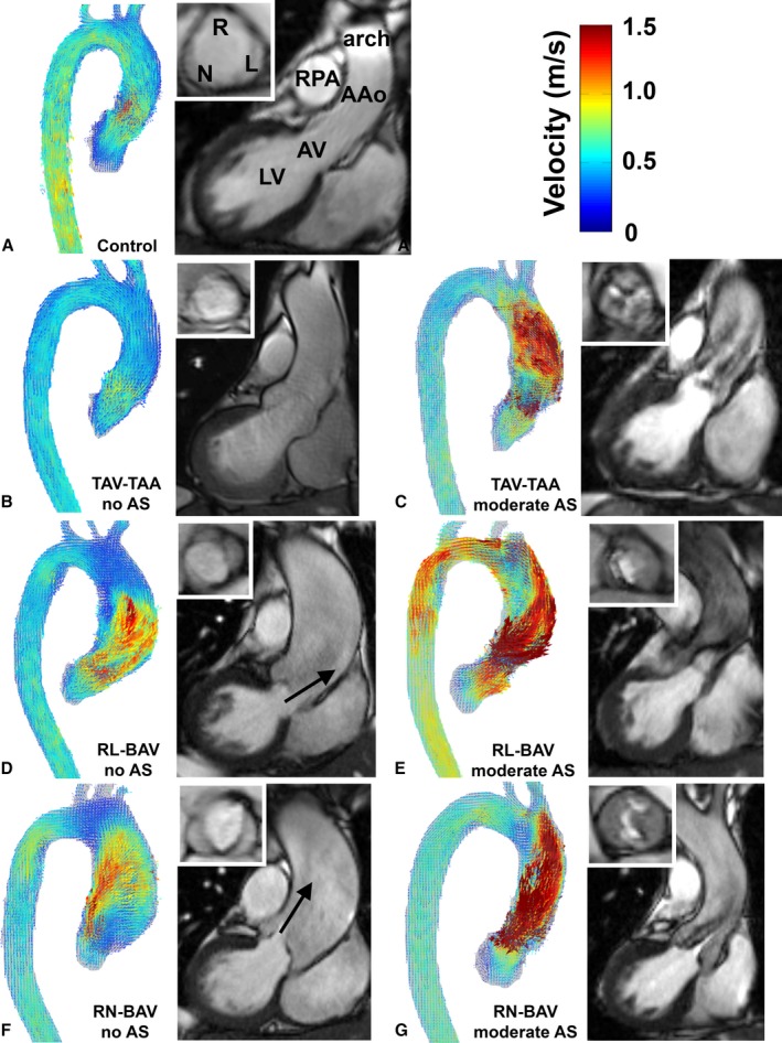
Figure 2.

Representative examples of systolic 3D velocity fields in the aorta obtained by 4D flow MRI, aortic valve morphology (white inset box) and left ventricular outflow based on 2D CINE SSFP MRI for (A) controls, (B) TAV‐TAA without AS, (C) TAV‐TAA with moderate AS, (D) RL‐BAV without AS, (E) RL‐BAV with moderate AS, (F) RN‐BAV without AS and (G) RN‐BAV with moderate AS. 2D indicates 2‐dimensional; 3D, 3‐dimensional; 4D, 4‐dimensional; AAo, ascending aorta; AS, aortic stenosis; AV, aortic valve; BAV, bicuspid aortic valve; LV, left ventricle; MRI, magnetic resonance imaging; RL‐BAV, right and left coronary leaflet fusion BAV; RN‐BAV, right and noncoronary leaflet fusion BAV; RPA, right pulmonary artery; SSFP, steady state free precession; TAV‐TAA, tricuspid aortic valve with aortic dilation. Arrows indicate high‐velocity outflow jets in RL‐BAV and RN‐BAV without AS.
