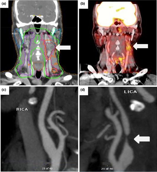
Figure 3.

A series of images from a patient positive for human papillomavirus (HPV) who had radiation therapy to the left side of the neck for hypopharyngeal cancer and developed neurological complications. A, Radiation dose map computed tomography images showing dose exposure fields with an arrow pointing to the highest exposure area, which overlaps the left carotid artery. B, Surveillance positron emission tomography images taken 3.5 years after radiation therapy demonstrate increased inflammation (white arrow) around the left carotid. Subsequently, the patient reported several episodes of transient vision loss in the left eye and was diagnosed with a transient ischemic attack; computed tomography angiogram images demonstrate a normal right carotid and critical atherosclerotic stenosis in the proximal left carotid (C and D, arrows) at the same site as the area of exposure. LICA indicates left internal carotid artery; RICA, right internal carotid artery.
