Figure 7.

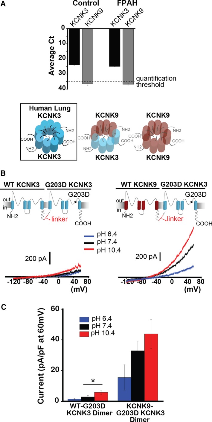
KCNK9 protects against KCNK3 dysfunction. A, Quantitative real‐time PCR analysis of human lung samples from healthy (Control) and familial PAH (FPAH) patient lungs. Expression of KCNK3 (black bars), and KCNK9 (gray bars) are compared, based on mean cycle threshold (Ct) values observed for each gene; Ct>35 indicates no quantifiable gene expression (n=5 patient lungs for each lane). Of the possible KCNK3+KCNK9 channel combinations, only KCNK3 homomeric channels (boxed, left) are predicted to form in human lungs. B, Voltage clamp recordings of the WT‐G203D KCNK3 heterodimer (left), and the KCNK9‐G203D KCNK3 heterodimer (right), showing current traces at pH 6.4 (blue), 7.4 (black), and 10.4 (red). C, Summary of current densities (pA/pF at 60 mV) at pH 6.4, 7.4, and 10.4 (n=4–14 cells per pH bar). Bar graphs show mean±SEM. *P<0.05 by the unpaired Student t test. PAH indicates pulmonary arterial hypertension; PCR, polymerase chain reaction; WT, wildtype.
