Figure 6.

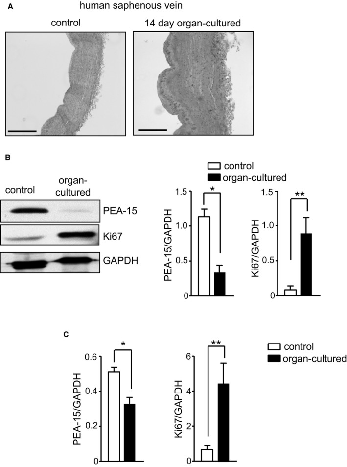
PEA‐15 (phosphoprotein enriched in astrocytes 15) expression is dynamically regulated in human arteries in an ex vivo model of neointimal hyperplasia. A, Representative image of human saphenous vein segments from the same patient, comparing the wall thickness of control and 14‐day organ‐cultured segments. Scale bar=400 μm. B, Immunoblots from control and organ‐cultured vein segments to determine expression of PEA‐15, Ki67 (proliferative marker), and GAPDH. Representative immunoblots are shown. Mean data for PEA‐15 and Ki67 protein expression are calculated as a ratio of GAPDH, n=6. *P<0.05 and **P<0.01 using Student t test. C, Gene expression of PEA‐15 and Ki67 in control and organ‐cultured human saphenous vein segments determined by quantatitive polymerase chain reaction. Results are expressed as a ratio of GAPDH, n=6. *P<0.05 and **P<0.01 using Student t test.
