
Figure 3.
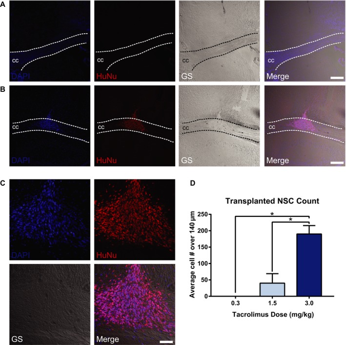
Immunosuppression requirement for CC‐targeted NSC survival in AD mice. Representative confocal imaging of NSC 4 weeks after CC targeting in Tg‐AD mice immunosuppressed with tacrolimus at 0.3 mg/kg (A) and 3.0 mg/kg (B). Immunosuppression with 0.3 mg/kg shows no evidence of surviving NSCs in the targeted area (A). HuNu‐positive NSCs are visible in the CC at the 3.0 mg/kg dose, with extensive NSC survival and migration along the white matter tract structure (B–C). NSCs are labeled with HuNu (red); nuclei are labeled with DAPI (blue); CC, corpus callosum; GS, grayscale. (A–C) scale bar 200 μm; D: scale bar 100 μm. Cell counts expressed as average numbers of HuNu‐positive cells over a distance of 140 μm within the injection coordinates (−0.8 to −1.6 mm from bregma) show significantly increased NSCs with 3.0 mg/kg tacrolimus versus other doses tested (D). (*P < 0.05).