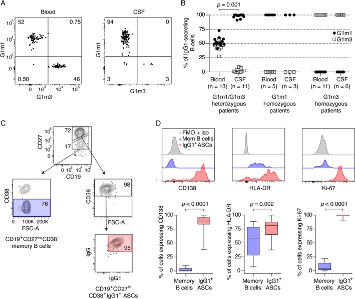
Figure 2.

Antibody‐secreting cells of the G1m1 allotype are selectively enriched in the cerebrospinal fluid (CSF) of G1m1/G1m3 heterozygous MS patients, and the cells have a phenotype compatible with highly proliferating plasmablasts. (A) A representative flow cytometry experiment from a G1m1/G1m3 heterozygous MS patient. The cells were gated for expression of IgG1 as shown in Fig. 1, and assayed for the expression of G1m1 and G1m3. (B) The frequencies of G1m1 and G1m3 antibody‐secreting cells in the CSF of G1m1/G1m3 heterozygous patients (left panel), G1m1 homozygous patients (middle panel) and G1m3 homozygous (right panel) are shown. Double negative cells (in CSF median [range] 0% [0–3.8%] and in blood 2.7% [1.4–7.2%]) were excluded when estimating the frequencies. Two G1m1/G1m3 heterozygous patients, two G1m1 homozygous patients, and five G1m3 homozygous patients had no detectable IgG1‐secreting B cells in the CSF. Frequencies of G1m1‐ and G1m3‐expressing cells were compared between CSF and blood for G1m1/G1m3 heterozygous patients (n = 11 paired samples, Wilcoxon signed‐rank test). (C) Antibody‐secreting cells (ASCs) were gated for the expression of CD19 and high levels of CD27, in addition to the expression of CD38, IgG, and IgG1, while memory B cells were gated for the expression of CD19, intermediate levels of CD27, and absence of CD38. (D) The frequencies of IgG1 ASCs and memory B cells expressing CD138, HLA‐DR, and the proliferation marker Ki‐67 (n = 20, Wilcoxon signed‐rank test). Positive gates were set using fluorescence minus one with isotype controls (FMO + iso). Results are presented as box plot diagrams with median, upper and lower quartile, and minimum/maximum values.
