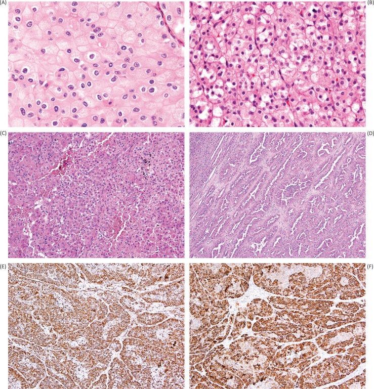
Figure 4.
Mimics of oncocytoma include eosinophilic variant chromophobe renal cell carcinoma (A), in which distinction from oncocytoma is challenging, but is favored by perinuclear clearing (“halo”), nuclear wrinkling and irregularity, and substantial trabecular growth pattern. Succinate dehydrogenase-deficient renal cell carcinoma (B) is a more recently recognized entity that may closely mimic oncocytoma due to bland, uniform cytology; however, clues to this diagnosis include diffuse growth pattern (rather than tubular or nested formation) and cytoplasmic inclusions of flocculent material (sometimes referred to as vacuoles), likely representing large abnormal mitochondria. Papillary renal cell carcinoma with oncocytic features (C) can also mimic oncocytoma; however, this diagnosis should be suspected in the presence of substantive papillary formation (D) or positivity for vimentin (E). Labeling for alpha-methyl-acyl-coA racemase (AMACR) is typically very strong and diffuse in all patterns of papillary renal cell carcinoma (F).

